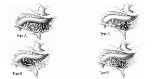
Direct carotid cavernous fistula
Direct carotid cavernous fistula (CCF) are high flow fistulas occurring due to a tear in the carotid artery most commonly from either penetrating or non-penetrating head trauma.
Direct CCF also occur secondary to cavernous aneurysm rupture and from iatrogenic trauma following oromaxillofacial and neurosurgical procedures.
The most common (70%-90%) etiology of direct CCF is trauma from a basal skull fracture resulting in tear in the internal carotid artery (ICA) within the cavernous sinus.
Videos
A video of Liao et al., from the Chung Shan Medical University, Institute of Medicine, Taichung. Departments of Neurosurgery and Department of Neuroradiology, Taichung Veterans General Hospital, Neurology, Neurological Institute, and Department of Neurosurgery, Tri-Service General Hospital, National Defense Medical Center, Taipei, Taiwan presents a case of new-onset visual blurring, diplopia, and conjunctival injection after head injury. CTA of the brain revealed a direct carotid-cavernous fistula (dCCF) of the right side. Careful evaluation of CTA source images revealed that the fistula point was at the ventromedial aspect of the right cavernous internal carotid artery (ICA), about 3.6 × 3.6 mm2 in size, with 3 main outflow channels (2 intracranial and 1 extracranial) (CTA-guided concept). DSA of the brain also confirmed the diagnosis but was unable to locate the fistula point in a large-sized dCCF. Through a transfemoral artery approach, 3 microcatheters were navigated to each peripheral channel to initiate outflow-targeted embolization. Intracranial refluxes were blocked first to avoid cerebral hemorrhages, followed by the extracranial outflow. During embolization, accidental dislodge of one coil into the sphenoparietal vein occurred, but no attempt of coil retrieval was made. Complete obliteration of the dCCF was achieved, and the patient recovered well without new neurological deficits. 4D MRA at the 3-month follow-up showed no residual dCCF.The video can be found here: https://youtu.be/LH2lNVRZSPk
<html><iframe width=“560” height=“315” src=“https://www.youtube.com/embed/LH2lNVRZSPk” frameborder=“0” allow=“accelerometer; autoplay; encrypted-media; gyroscope; picture-in-picture” allowfullscreen></iframe> </html> 1).
Case reports
A 58-year-old woman with symptoms of blepharoptosis and intracranial bruits for 1 wk. During physical examination, there was right eye exophthalmos and ocular motor palsy. The rest of the neurological examination was clear. Notably, the patient had no history of head injury. The patient was treated with a pipeline embolization device in the ipsilateral internal carotid artery across the fistula. Coils and Onyx were placed through the femoral venous route, followed by placement of the pipeline embolization device with assistance from a balloon-coiling technique. No intraoperative or perioperative complications occurred. Preoperative symptoms of bulbar hyperemia and bruits subsided immediately after the operation.
Conclusion: Pipeline embolization device in conjunction with coiling and Onyx may be a safe and effective approach for direct CCFs 2).
