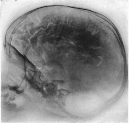
Pneumoencephalography
Pneumoencephalography (sometimes abbreviated PEG; also referred to as an “air study”) was a common medical procedure in which most of the cerebrospinal fluid (CSF) was drained from around the brain by means of a lumbar puncture and replaced with air, oxygen, or helium to allow the structure of the brain to show up more clearly on an X-ray image. It was derived from ventriculography, an earlier and more primitive method where the air is injected through holes drilled in the skull.
The procedure was introduced in 1919 by the American neurosurgeon Walter Edward Dandy and was performed extensively until the late-1970s when it was replaced by more sophisticated and less-invasive modern neuroimaging techniques.
In the era before computerized tomography (CT), extradural hematomas were usually diagnosed by invasive and less accurate techniques, such as cerebral angiography, pneumoencephalography, or exploratory burr holes.
In his serially published atlas of pathology, Anatomie Pathologique du Corps Humain (1829-1842), French anatomist and pathologist Jean Cruveilhier (1791-1874) provided an early clinical-pathologic description of Dyke-Davidoff-Masson syndrome. Cruveilhier's case was initially published around 1830, more than a century before the clinical and radiologic report of Dyke and colleagues in 1933 based on a series of patients studied with pneumoencephalography. Although Dyke and colleagues were apparently unaware of Cruveilhier's prior description, Cruveilhier's case manifested all of the key osseous and neuropathological features of Dyke-Davidoff-Masson syndrome as later elaborated by Dyke and colleagues: (1) cerebral hemiatrophy with ex vacuo dilation of the lateral ventricle, (2) ipsilateral thickening of the diploe of the skull, and (3) ipsilateral hyper-pneumatization of the frontal sinuses. In addition, Cruveilhier noted crossed cerebral-cerebellar atrophy in his case and correctly inferred a “crossed effect” between the involved cerebral hemisphere and the contralateral cerebellum. Cruveilhier's pathological case from 1830 clearly anticipated both the cases reported more than a century later by Dyke and colleagues based on pneumoencephalography and the more recent case reports recognized with computed tomography or magnetic resonance imaging 1).
