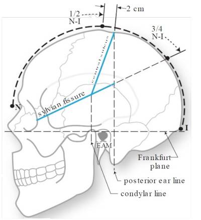
Sylvian fissure
The lateral sulcus also called Sylvian fissure (SyF) or lateral fissure is one of the most prominent structures of the brain (the fissure separating the temporal lobe from the parietal lobe and frontal lobes).
The central sulcus joins the Sylvian fissure in only 2 % of cases.
The Sylvian fissure terminates in the supramarginal gyrus (Brodmann area 40).
Approximated by a line connecting the lateral canthus to the point 3/4 of the way posterior along the arc running over convexity from nasion to inion (T-H lines).
The frontotemporal, so-called pterional approach has evolved with the contribution of many neurosurgeons over the past century. It has stood the test of time and has been the most commonly used transcranial approach in neurosurgery. In its current form, drilling the sphenoid wingas far down as the superior orbital fissure with or without the removal of the anterior clinoid process, thinning the orbital roof, and opening the Sylvian fissure and basal cisterns are the hallmarks of this approach.
The bone flap has been removed and the dura mater has been opened as a flap pediculated towards the greater sphenoid wing previously roungered to improve parasellar visualization. Sylvian fissure, Inferior frontal gyrus, Superior temporal gyrus and Middle temporal gyrus are exposed. Three pars of parasylvian inferior frontal gyrus must be distinguished: pars orbitalis (pOr) in relation to the orbital roof; pars triangularis (pT) the widest area of sylvian fissure (good place for start opening of sylvian fissure); pars opercularis (pOp) where Broca’s Area is located.
Compartments
The sylvian fissure extends from the basal to the lateral surface of the brain and presents 2 compartments on each surface,
1 superficial (temporal stem and its ramii) and 1 deep (anterior and lateral operculoinsular compartments). The temporal operculum is in opposition to the frontal and parietal opercula (planum polare versus inferior frontal and precentral gyri, Heschl's versus postcentral gyri, planum temporale versus supramarginal gyrus). The inferior frontal, precentral, and postcentral gyri cover the anterior, middle, and posterior thirds of the lateral surface of the insula, respectively. The pars triangularis covers the apex of the insula, located immediately distal to the genu of the middle cerebral artery. The clinical application of the anatomic information presented in the article of Wen et al. is in angiography, middle cerebral artery aneurysm surgery, insular resection, frontobasal resection, and amygdalohippocampectomy, and hemispherotomy 1).
Divisions
The SyF is divided into a proximal segment and a distal segment separated by the anterior sylvian point (ASP).
It is the single most identifiable feature of the superolateral face of the brain, and together with the underlying sylvian cistern it constitutes the most frequently used microneurosurgical corridor because of the high proportion of intracranial lesions that are accessible through its opening 2).
The lateral sulcus divides both the frontal lobe and parietal lobe above from the temporal lobe below. It is in both hemispheres of the brain but is longer in the left hemisphere in most people. The lateral sulcus is one of the earliest-developing sulci of the human brain. It first appears around the fourteenth gestational week.
The sylvian fissure or lateral sulcus is the most identifiable feature of the superolateral brain surface and constitutes the main microneurosurgical corridor, given the high frequency of approachable intracranial lesions through this route.
In their original description of the microsurgical anatomy of the subarachnoid cisterns in 1976, Yasargil, et al., 3) emphasized the importance of the SyF, which then became the main microneurosurgical corridor to the base of the brain. In later publications Yasargil, et al., described in detail the microanatomy of this fissure and its underlying cistern 4) 5) 6) and the technique of its opening.
The opening of the fissure at the level of the anterior sylvian point (ASP) shows very soon the insular apex. The limen insula and the middle cerebral artery bifurcation are a little bit deeper and 1-2 cm. anteriorly. The opening of the sylvan fissure posteriorly to the ASP exposes the insula and the opening anteriorly leads to the suprasellar cisterns. The distance between the ASP and the IRP along the SF is 2.3 cm.
Branches
The lateral sulcus has a number of side branches. Two of the most prominent and most regularly found are the ascending (also called vertical) ramus and the horizontal ramus of the lateral fissure, which subdivide the inferior frontal gyrus. The lateral sulcus also contains the transverse temporal gyri, which are part of the primary and below the surface auditory cortex.
Partly due to a phenomenon called Yakovlevian torque, the lateral sulcus is often longer and less curved on the left hemisphere than on the right.
It is also located near Sylvian Point.
The area lying around the Sylvian fissure is often referred to as the perisylvian cortex. The human secondary somatosensory cortex (S2, SII) is a functionally-defined region of cortex in the parietal operculum on the ceiling of the lateral sulcus.
Yasargil divides the SyF into a proximal segment (stem, sphenoidal, anterior ramus) and a distal segment (lateral, posterior ramus) separated by the sylvian point 7) 8). which is located beneath the triangular part of the inferior frontal gyrus (IFG).
The horizontal and the anterior ascending branches of the SyF that delineate the triangular part of the IFG arise at the sylvian point 9).
Variants
Yasargil described four different types of intraoperatively observed anatomical sylvian fissure (SF) variants.
Category I is a straight wide SF, II a straight narrow SF, III a herniated frontal lobe into the SF and IV is a herniated temporal lobe into the SF. 10).
The SF categories used in the present work are based on the Yasargil classification with slight modifications since we categorized the SF on cranial computed tomography (CCT) scans and not anatomically.
Splitting
Sylvian fissure dissection is an essential microneurosurgical skill for neurosurgeons. The safe and accurate opening of the Sylvian fissure is desirable for a good prognosis.
Insular gliomas represent a unique surgical challenge due to the complex anatomy and nearby vascular elements associated within the Sylvian fissure. For certain tumors, the transsylvian approach provides an effective technique for achieving maximal safe resection.
The goal of the manuscript and video of Safaee et al., are to present and discuss the surgical nuances and appropriate application of splitting the Sylvian fissure. The hope is that this video highlights the safety and efficacy of the transsylvian approach for appropriately selected insular gliomas 11).
Pathology
Development
After sagittal division of the prosencephalon at 4.5 weeks of gestation, the early fetal cerebral hemisphere bends or rotates posteroventrally from seven weeks of gestation. The posterior pole of the telencephalon thus becomes not the occipital lobe but the temporal lobe as the telencephalic flexure forms the operculum and finally the lateral cerebral or Sylvian fissure 12).


